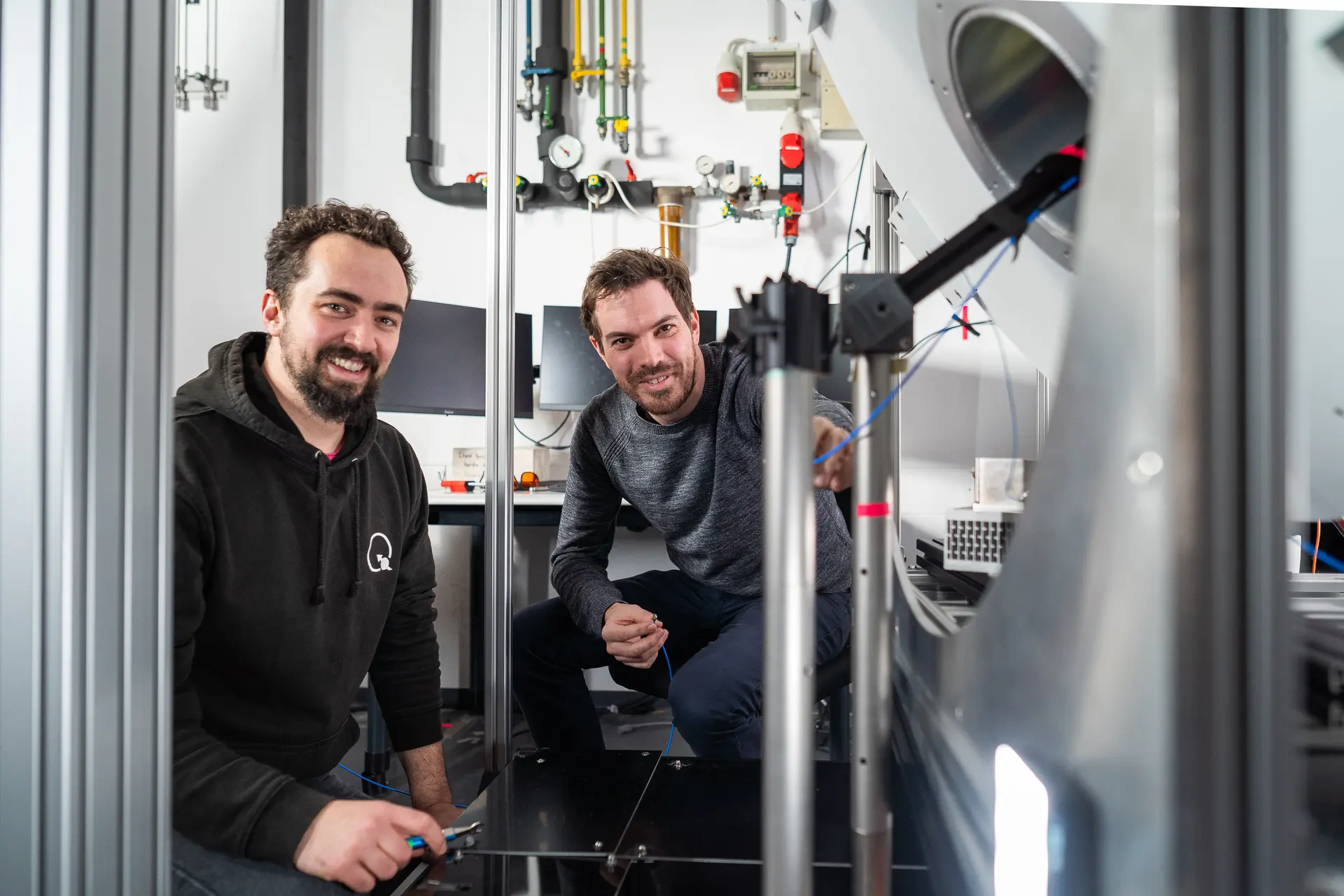
Researchers at the Technical University of Munich (TUM) have invented an entirely new field of microscopy, nuclear spin microscopy. The team can visualize magnetic signals of nuclear magnetic resonance with a microscope. Quantum sensors convert the signals into light, enabling extremely high-resolution optical imaging.
Magnetic resonance imaging (MRI) scanners are known for their ability to look deep into the human body and create images of organs and tissues. The new method, published in the journal Nature Communications, extends this technique to the realm of microscopic detail. "The quantum sensors used make it possible to convert magnetic resonance signals into optical signals. These signals are captured by a camera and displayed as images," explains Dominik Bucher, Professor for Quantum Sensing and researcher at the Cluster of Excellence Munich Center for Quantum Science and Technology (MCQST).
Publication
Karl D. Briegel, Nick R. von Grafenstein, Julia C. Draeger, Peter Blümler, Robin D. Allert & Dominik B. Bucher: Optical widefield nuclear magnetic resonance microscopy, published in: Nature Communications, February 03, 2025, https://doi.org/10.1038/s41467-024-55003-5
Further information and links
- The start-up team QTAS has already been formed based on the new technology. Using magnetic resonance microscopy, it aims to detect individual cancer cells with high precision and at short intervals. The team has received the Medical Valley Award 2024 from the Bavarian Ministry of Economic Affairs.
- The research project was funded by the Bavarian State Ministry of Science and the Arts as part of the IQSense project via Munich Quantum Valley (MQV), by the Federal Ministry of Education and Research (BMBF) as part of the VIP+ Validation funding program and the Munich Center for Quantum Science and Technology (MCQST).
- Quantum science at TUM
Original Article: https://www.tum.de/en/news-and-events/all-news/press-releases/details/a-completely-new-type-of-microscopy-based-on-quantum-sensors